A gallbladder polyp on a scan does not automatically mean cancer or an operation. Size, shape, wall changes, previous scans and individual risks guide the next step.
An ultrasound done for another reason mentions a small gallbladder polyp. You may have no pain, but the unfamiliar word raises two immediate questions: is this a stone, and does it need an operation?
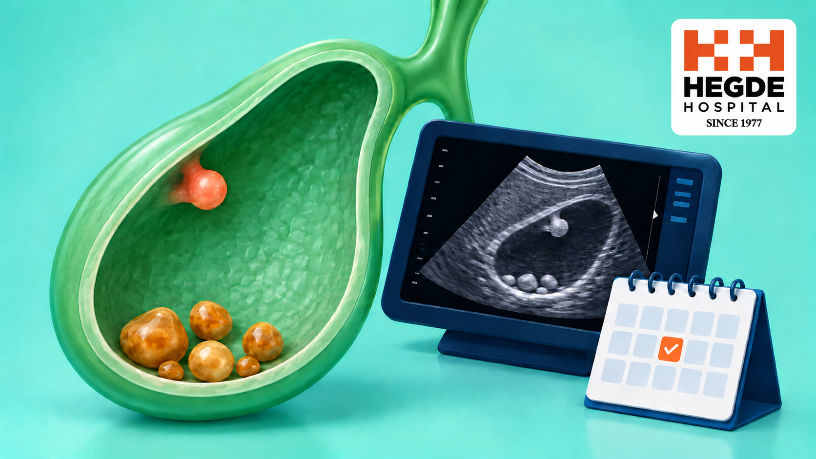
A scan finding needs interpretation in context. A polyp is an area projecting from the inner gallbladder wall. Many small findings are benign, including cholesterol deposits that resemble polyps. The report alone may not identify the exact tissue type, so the next decision involves both the images and your health history.
The direct answer
Gallbladder polyps and gallstones are different findings. Some small, reassuring polyps can be observed, while larger or suspicious findings need a surgical opinion. The largest measured size, shape, adjacent wall thickening, change on comparable scans and individual risk factors matter together. There is no single follow-up timetable suitable for every patient.
How is a polyp different from a stone or sludge?
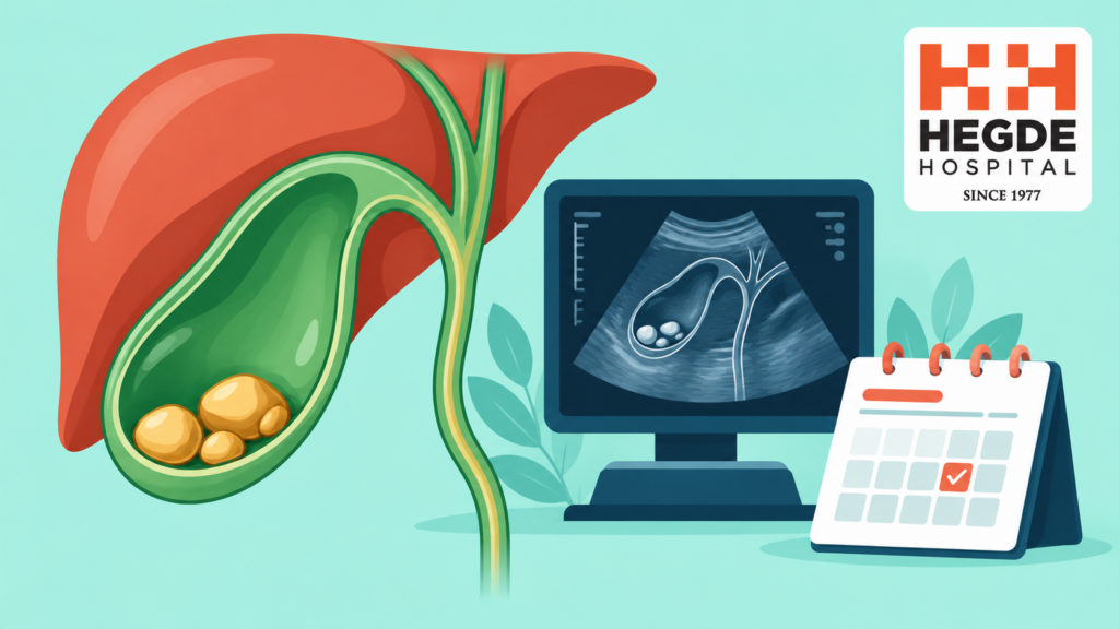
The gallbladder stores bile made by the liver. A polyp-like finding is attached to its inner wall; a stone is hardened material within the bile. Sludge is thickened material that can sometimes resemble a wall lesion. Ultrasound helps distinguish them, although the distinction is not always certain on one examination.
| Finding | Typical scan distinction | What it means for the discussion |
|---|---|---|
| Polyp-like projection | Arises from or appears attached to the wall | Review size, shape and the surrounding wall |
| Gallstone | Often moves with position and may cast a shadow | Follow the separate stone-related assessment |
| Sludge or an uncertain finding | May imitate a polyp on an initial scan | A radiologist may clarify the finding with repeat or selected imaging |
These are usual patterns, not instructions for reading your own images. Stones and polyps can also occur together. The gallstone symptom guide explains a different clinical pathway.
Which details should you look for in the report?
Check whether the largest finding is measured in millimetres and whether earlier images are available. A measurement of 5 mm is not the same as 5 cm. If there are several polyps, the largest one usually guides size-based decisions, but the overall appearance still matters.
The report may describe a narrow stalk or a broad attachment, also called a sessile appearance. Nearby wall thickening and an uncertain or suspicious appearance can change the plan. A missing description is a reason to ask for clarification, not proof of a dangerous lesion.
Tell the clinician about age, relevant previous illness and any diagnosed bile-duct condition, particularly primary sclerosing cholangitis. Some guidance also considers ethnicity when discussing risk. An Indian patient should have an individualized review rather than apply a monitoring chart from another population without checking that it fits.
Does a polyp mean cancer?
No. Many small gallbladder polyps are non-cancerous. However, ultrasound cannot always determine tissue type with certainty, and a larger or concerning lesion deserves specialist assessment. Neither a small size nor absence of pain provides an absolute guarantee.
Avoid judging the risk from the number of polyps alone. One polyp is not automatically cancer, and several are not automatically harmless. The clinician combines the imaging features, size, history and any change over time.
When is monitoring reasonable, and when is surgery discussed?
For an appropriate small finding with reassuring features, a clinician may recommend observation with a defined ultrasound plan. Some very small low-risk findings may not need ongoing surveillance. The decision should state when to return and what change would trigger reassessment.
A finding around 10 mm or larger is an important reason to discuss the report with a specialist. Professional guidelines differ: some consider surgery from 10 mm, while others use higher thresholds for reassuring appearances and lower thresholds for concerning features. Size alone is therefore not a universal operation rule.
Planned gallbladder removal may be considered when the expected benefit outweighs surgical risks. Suspicious wall changes, growth assessed in context or relevant personal risks can influence that discussion. If an operation is proposed, ask why observation is less suitable for your particular finding.
The usual operation removes the gallbladder, rather than simply plucking off the polyp. The removed tissue can then be examined. The surgical approach, recovery and need for further care depend on the findings and your overall health; there is no guaranteed recovery date for everyone.
What if the measurement has increased slightly?
A small difference between scans can reflect measurement variation or benign growth. The clinician should compare the actual images, dates, largest dimension and appearance before deciding that a change is significant. A single additional millimetre is not itself a cancer diagnosis.
Bring prior reports and images, preferably from all relevant examinations. If a repeat study is advised, ask when, where and whether any preparation is required. More detailed ultrasound or other imaging is reserved for selected uncertain cases; every patient does not need CT, MRI and an endoscopic examination.
Can the polyp explain pain, and can food remove it?
A polyp found during a pain investigation may be incidental. Stones or other digestive conditions may explain symptoms more convincingly. An operation directed at the polyp does not guarantee that unrelated bloating or discomfort will disappear.
Food changes do not reliably remove a gallbladder polyp. Do not use a diet claim as a reason to abandon recommended review. Advice about gallstones and food choices addresses a separate issue and does not establish a polyp treatment.
How soon should you arrange review?
A stable incidental finding without concerning symptoms usually allows a planned appointment. Arrange an earlier review if the report describes a suspicious lesion or if discomfort is recurring or worsening. Severe persistent abdominal pain, fever or yellowing of the eyes requires urgent qualified medical assessment.
At the consultation, ask: is this confidently a polyp, what is the largest size, are there concerning wall features, and what is my written follow-up plan? Bring the full report rather than an isolated measurement. You can view Hegde Hospital doctors or request an appointment to discuss the appropriate specialist.
Frequently asked questions
Does a 4 or 5 mm gallbladder polyp always need surgery?
No. A small finding often allows observation or, in selected low-risk circumstances, no continuing scans. The appearance and personal risk factors still need review before deciding the plan.
Is a 10 mm polyp definitely cancer?
No. Ten millimetres is a size that commonly prompts a more detailed surgical discussion. It is not a tissue diagnosis, and recommendations depend on the guideline, imaging features and individual circumstances.
If I have no pain, can I ignore the finding?
Absence of pain does not determine the whole plan. Have the report reviewed once, then follow the agreed schedule or documented decision that further surveillance is unnecessary.
Can a polyp disappear on the next ultrasound?
Some initially reported polyps are deposits, sludge or findings that are not reproduced later. A clinician should confirm what the new scan means and whether monitoring can stop; do not make that decision from symptoms alone.
Should every scan change lead to gallbladder removal?
No. The size difference, scan comparability, time interval and any new suspicious features need to be assessed together. Ask the specialist to explain why continued observation or planned surgery fits your situation.